Short answer
An otoscope is a hand-held instrument that lights and magnifies the ear canal and eardrum. An ophthalmoscope is used to examine the fundus of the eye (retina, optic disc and vessels), and a negatoscope (X-ray film viewer) lights radiographs evenly from behind so they can be read. With the wooden tongue depressor, they form an exam room's core diagnostic kit.
What is an otoscope and what is it used for?
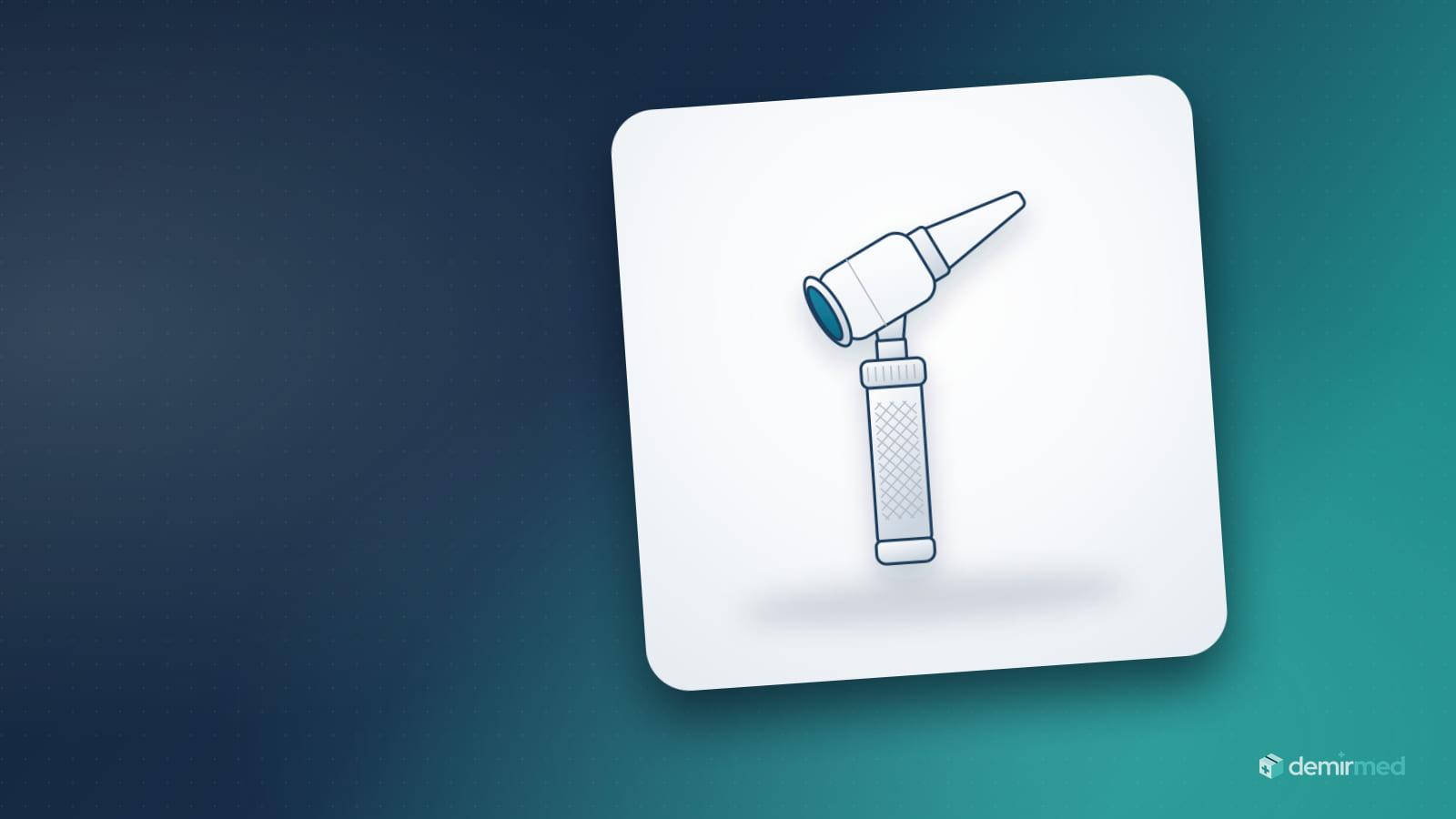
The name joins the Greek oto- (ear) and -scope (to look): literally, an instrument for looking into the ear. In otoscopic examination the clinician assesses the skin of the ear canal, wax, any foreign body, and the colour, light reflex and integrity of the eardrum (tympanic membrane); impacted wax, otitis externa and signs of otitis media are all seen this way. It is the most frequently used ear examination instrument in family medicine, ENT, paediatrics, emergency departments and school and occupational health units. Hand-held models are usually boxed with a reusable speculum set.
Parts of an otoscope
An otoscope consists of a handle and a head; the quality of the examination depends on the optics, light source and speculum on the head.
| Part | Function | What to check when buying |
|---|---|---|
| Handle | Holds the batteries or rechargeable cell; on most models a rotating ring on top (rheostat) switches the light on and adjusts it | 2.5 V battery or 3.5 V rechargeable; match with the head |
| Head | Holds the light source, viewing window and speculum mount | Threaded or bayonet fitting |
| Magnifying viewing window | Magnifies the image about 3x on most models (roughly 2.5–4x by model) | Window that opens for passing instruments |
| Light source | Illuminates the ear canal | Halogen/xenon or LED; direct or fibre-optic delivery |
| Speculum (otoscope tip) | Cone that sits in the ear canal and forms the viewing channel | Tip diameter, single-use or reusable, fit with the head |
| Insufflation port | A rubber bulb is attached for pneumatic otoscopy | Sealed closed head |
Otoscope light source: halogen, fibre-optic and LED
The light is produced by a halogen/xenon bulb or an LED, and it reaches the examination area either directly or through fibre optics. With conventional (direct) illumination the bulb sits inside the head; it can block part of the view and cause reflections. In a fibre-optic system the light is carried to the tip by fine fibres arranged around the speculum opening; the view stays unobstructed and the tip gives a ring of light without noticeable heat.
| Feature | Halogen / xenon (direct) | Fibre-optic delivery | LED |
|---|---|---|---|
| Light produced by | Bulb in the head | Bulb or LED; light carried to the tip by fibres | Semiconductor diode |
| Colour temperature (approx.) | 2,700–3,300 K, warm yellowish white; xenon-halogen slightly whiter | Depends on the source | Mostly 3,000–4,500 K by model; cooler white models exist |
| Heat | Bulb warms up | No heat felt at the tip | Very low |
| Field of view | Bulb and reflections may partly affect the view | Unobstructed ring light | Used in direct or fibre-optic designs |
| Life and maintenance | Limited bulb life; spare bulbs needed | Depends on the source; broken fibres reduce light | Manufacturers quote tens of thousands of hours; on most models no replacement is needed |
| Power use | High; batteries run down faster | Depends on the source | Low; batteries last longer |
Because the colour of the eardrum carries diagnostic information, an LED should also be chosen for colour fidelity (high CRI). When buying, ask for the light source of the model (LED or halogen) to be stated in the quotation.
Otoscope tip (ear speculum) sizes and hygiene
The ear speculum, often simply called the otoscope tip, is specified by its tip diameter in millimetres. Reusable sets on the market most often contain 2.5, 3, 4 and 5 mm tips; some sets also include smaller tips such as 2 mm. Single-use tips are usually made in two sizes: about 2.5–3 mm paediatric and 4–4.5 mm adult. Values vary by manufacturer.
| Tip diameter (approx.) | Typical use |
|---|---|
| 2–2.5 mm | Newborns and infants, very narrow canals |
| 3 mm | Young children |
| 4 mm | Older children, narrow adult canals |
| 5 mm | Wide adult canals |
The general rule is to choose the largest diameter that sits comfortably in the ear canal: a wider speculum gives more light and a wider view, while one that is too small narrows the view and tempts deeper insertion.
- Reusable specula: Plastic or metal; after each patient they are first cleaned and then disinfected according to the manufacturer's instructions for use (IFU). They reduce waste but need reprocessing time and spares to rotate.
- Single-use specula: A new tip is fitted for each patient and discarded after use; because they reduce cross-contamination risk and reprocessing workload, they are practical in busy clinics and emergency departments. Tips are often specific to the head model, so check compatibility before buying, and never wash and reuse them.
Ask for the contents of the speculum set supplied with the device (number and diameters) to be stated in the quotation. If you plan to use single-use tips, specify the device model and plan compatible tips alongside it.
The basic principle of ear examination
The external ear canal is slightly S-shaped; to straighten it, the auricle is gently pulled up and back in adults and down and back in young children. The speculum is placed only in the outer (cartilaginous) part of the canal; the inner bony part is very sensitive. Interpretation of findings and treatment decisions rest with the physician and local protocol.
Pneumatic otoscopy and the video otoscope
In pneumatic otoscopy a rubber bulb is attached to the insufflation port of a closed head; with a speculum that seals the ear canal, a slight pressure change is applied and the mobility of the eardrum is observed. When fluid is present in the middle ear, eardrum movement is reduced.
In a video otoscope a camera in the head sends the image to a screen or computer; recording is useful for teaching and remote consultation. Recordings are sensitive personal health data and must be stored in line with data protection law.
What is an ophthalmoscope? Direct versus indirect ophthalmoscopes
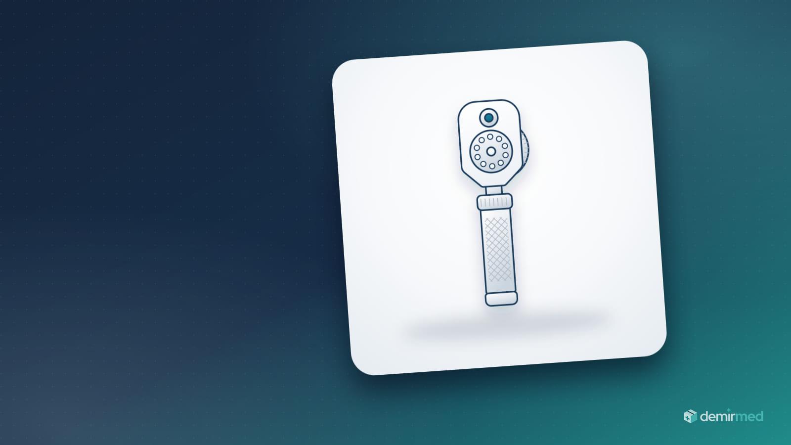
An ophthalmoscope sends light through the pupil so the examiner can see the fundus of the eye. In Turkish it is spelled "oftalmoskop", although "oftalmaskop" is a common variant. There are two main types: the hand-held direct ophthalmoscope and the indirect ophthalmoscope, which uses a head-mounted light source and a hand-held lens.
| Feature | Direct ophthalmoscope | Indirect ophthalmoscope |
|---|---|---|
| Design | Hand-held; handle and head | Head-mounted binocular light source and hand-held condensing lens (often 20 D) |
| Image | Upright | Inverted and laterally reversed |
| Magnification (approx.) | 15x | About 3x with a 20 D lens |
| Field of view (approx.) | Narrow, 5–10 degrees | Wide, about 45 degrees with a 20 D lens |
| Depth perception | Monocular; not stereoscopic | Binocular; stereoscopic |
| Pupil | Usable through a small pupil; a dilated pupil makes the view easier | Usually needs a dilated pupil |
| Main users | Family medicine, internal medicine, neurology, emergency, paediatrics | Mainly ophthalmologists |
Direct ophthalmoscope features: lens wheel, apertures and filters
The values below are typical and vary by model.
| Feature | What it does | Typical value or availability |
|---|---|---|
| Lens wheel (Rekoss disc) | Compensates for the refractive error of examiner and patient to bring the fundus into focus | About −20 to +20 D on basic models, about −30 to +40 D or wider on advanced ones; mostly in 1 D steps. On most models red figures mark minus lenses and black or green figures mark plus lenses |
| Small spot | Reduces corneal reflection through a small (undilated) pupil | Standard |
| Large spot | Widest fundus view through a dilated pupil | Standard |
| Micro spot or half-moon | Viewing through very small pupils and reducing reflection | Some models |
| Slit | Helps tell raised from depressed lesions and roughly estimate anterior chamber depth | Most models |
| Fixation target or grid | Assessing fixation; roughly measuring lesion size and distance from the optic disc | Most models |
| Red-free (green) filter | Makes vessels, haemorrhages and the nerve fibre layer stand out; red lesions appear dark | Most models |
| Cobalt blue filter | Used with fluorescein staining to assess corneal abrasions and foreign bodies | Some models |
| Polarising filter | Reduces corneal reflections | Some models |
Hand-held direct ophthalmoscopes are focused with a dioptre lens wheel. The lens wheel range, aperture and filter options vary by model; when buying, ask for these features and the light source (LED or halogen) to be stated in writing in the quotation.
What is an ophthalmoscope used for?
- Fundus examination: Assessing the retina, macula and vessels; monitoring retinal changes related to hypertension and diabetes.
- Optic disc assessment: Disc margins, colour and cup-to-disc ratio; one of the first things checked when optic disc swelling (papilloedema) suggesting raised intracranial pressure is suspected.
- Red reflex screening: The red reflex examination in newborns and children is a screening tool for serious conditions such as cataract and retinoblastoma.
- Transparent media: Opacities in the cornea, lens and vitreous; a rough look at the anterior segment with a high plus lens.
The decision to dilate the pupil (mydriatic drops) is made by the physician, taking into account neurological observation, angle-closure risk and local protocol. Light intensity and examination time should be kept no higher or longer than needed; starting with low light improves patient comfort.
Otoscope
Otoscope parts and light source
Otoscope and ophthalmoscope sets: shared handle, voltage and charging
A diagnostic set consists of interchangeable heads that fit one handle. The most common combination is an otoscope and an ophthalmoscope; some sets include extra heads such as a throat illuminator. When buying a set, check:
- Handle and voltage: 2.5 V handles usually run on two alkaline batteries (C or AA size, by model); 3.5 V handles use a rechargeable cell (Li-ion or NiMH) and give a brighter, steadier light. The head and bulb must match the handle voltage.
- Charging options: Handles that plug into a socket, desk or wall chargers; wall-mounted diagnostic systems with two corded handles are also on the market.
- Rheostat: The rotating ring at the top of the handle switches the light on and adjusts it steplessly.
- Interchangeable heads: Threaded or bayonet fittings; handles and heads from different brands are not always compatible.
- Case: A case with compartments for heads, specula and spare bulbs protects the instruments from knocks and dust.
Otoscopes and ophthalmoscopes are sold both as separate devices and as sets sharing one handle; on both, the power source is in the handle, and the battery or rechargeable option should be confirmed in the quotation.
The laryngoscope: the same handle principle, a different purpose
A laryngoscope also consists of an illuminated handle and an attached blade; the light comes on when the blade is opened. It is not an examination-room device, however, but airway equipment for viewing the larynx during intubation, and its blade, which touches mucosa, needs high-level disinfection or sterilisation. For blade types and sizes, see our guide what is a laryngoscope, blade types and intubation.
Ophthalmoscope
Direct ophthalmoscope: lens wheel and filters
What is a negatoscope? Choosing an X-ray film viewer
A negatoscope is an illuminated panel that lights X-ray films evenly from behind so they can be read. Although digital imaging and PACS have spread, it is still needed wherever printed film is used, for comparing older films that patients bring, and in orthopaedics, chest medicine, dentistry, emergency departments and film archives. Thin, flicker-free, energy-saving LED viewers have replaced the old fluorescent-tube models.
| Criterion | What to look at | Practical note |
|---|---|---|
| Number of panels | Single or double; triple and quadruple models are also on the market | A double viewer lets two radiographs (AP and lateral, old and new) be read side by side |
| Viewing area | The largest film format you use | The most common large format is 35x43 cm; 24x30 and 18x24 cm films and dental films are smaller |
| Brightness | Value in cd/m² (nits) | A few thousand cd/m² is common for general radiography; dense films (e.g. mammography) need higher brightness |
| Uniformity | Brightness difference between centre and corners | Ask the manufacturer for it as a percentage; a patchy or dark-edged panel makes reading harder |
| Dimming | Stepless brightness control | For different film densities and ambient light |
| Film holder | Spring or roller clips | One-handed loading and holding several films |
| Mounting | Wall bracket or desk feet | Depends on room layout and reflections from windows and ceiling lights |
| Power and heat | Mains powered; consumption in watts | LED uses less energy and runs cooler than fluorescent models |
| Flicker-free light and colour | Constant-current LED driver, white close to daylight | Reduces eye fatigue during long reading sessions |
During reading, room light should be dimmed and windows and ceiling lights should not cause glare on the panel. Because the bright empty area around a small film dazzles the reader, some models have masking shutters.
Current LED viewers come in single and double versions, hold the radiograph with a film holder and run on mains electricity. When buying, ask for the dimensions and mounting (wall or desktop) to be confirmed in the quotation.
What is a tongue depressor (abeslang)? Use and storage
The wooden tongue depressor, called abeslang in Turkish (from the French abaisse-langue, "tongue lowerer"), is a single-use wooden blade that presses the tongue down during oral and throat examination so the inside of the mouth, the tonsils and the pharynx can be seen. It is also used to hold the tongue down while a throat swab is taken.
- Product design: Our wooden tongue depressor is a flat wooden blade with rounded ends, in standard adult length, with 100 pieces per box. Pack format (bulk or individually wrapped) depends on the lot, and the sterility status is printed on the label.
- Single use: A new depressor is used for each patient; being wood, it cannot be cleaned and reused, and after use it is discarded according to medical waste rules. Do not use a blade that is broken, splintered or damp.
- Storage: Keep boxes in a dry, clean, closed cupboard, off the floor and away from sink splashes; moisture can warp wood and cause mould. Never reach into an open box with used gloves.
- Stock: Multiply daily throat examinations by working days; a room doing 25 throat examinations a day uses a box of 100 in about four working days.
Paediatric-size, flavoured and plastic tongue depressors are also on the market; the item in our catalogue is a standard adult-length wooden depressor. For other examination consumables, see the Diagnostics & Test Products category.
X-ray viewer
Choosing an LED X-ray film viewer
Cleaning and infection control for examination devices
Reprocessing of medical devices follows the Spaulding classification: instruments entering sterile tissue are critical, those contacting mucosa or non-intact skin are semi-critical, and those touching only intact skin are non-critical. The bodies of examination devices are non-critical; parts that touch mucosa, such as a laryngoscope blade, are semi-critical. The exact method is set by the manufacturer's instructions for use (IFU) and the institution's infection control protocol; perform hand hygiene before and after patient contact.
| Device or part | When | Method (per IFU) | Avoid |
|---|---|---|---|
| Otoscope and ophthalmoscope handle, outer head surfaces | Between patients and at the end of the day | Wipe with a suitable surface disinfectant on a cloth or ready-made wipe | Immersing, spraying into openings, autoclaving unless the IFU allows it |
| Reusable speculum | After each patient | Clean first, then disinfect or sterilise as stated in the IFU; store dry in a closed container | Putting into disinfectant without cleaning |
| Single-use speculum and tongue depressor | Every patient | Discard into medical waste after use | Washing and reusing |
| Viewing window and optics | When soiled | Lens tissue or microfibre optical cloth | Abrasive cloths, paper towels, solvents that damage coatings |
| Laryngoscope blade | After every use | High-level disinfection or sterilisation; the handle is processed separately | Processing blade and handle the same way |
| Negatoscope screen and housing | Daily and when soiled | Switch off and unplug; soft cloth and a non-abrasive cleaner suitable for plastic | Abrasive powders or creams, solvents such as acetone, spraying liquid directly on the screen |
| Pulse oximeter | Between patients | Wipe the finger chamber and housing | Immersing |
| Couch mattress | Every patient | Change the cover or underpad, wipe the mattress | Using the same cover for the next patient |
The disinfectant must stay wet on the surface for the contact time on its label; bring the device to the patient's ear or eye only after it has dried completely.
Common otoscope, ophthalmoscope and negatoscope problems and fixes
| Problem | Likely cause | What to do |
|---|---|---|
| Dim otoscope or ophthalmoscope light | Weak battery, flat or ageing rechargeable cell, rheostat turned down, dirty contacts, blackened halogen bulb | Replace the battery or charge the handle, check the rheostat, clean the contacts per IFU, replace the bulb; if an LED model stays dim, send it for service |
| Light flickers or cuts out | Head not fully seated, loose bulb | Refit the head, check that the bulb is seated |
| Lens fogs up | Moving from a cold place to a warm room, damp storage | Let the device reach room temperature, wipe with an optical cloth, store in a dry case |
| Blurred ophthalmoscope image | Lens wheel not set for refractive error, dirty window | Turn the wheel until the image is sharp, clean the window with lens tissue |
| Corneal reflection hides the view | Large spot through a small pupil | Choose the small spot or, if available, the polarising filter |
| Ear canal not visible, patient uncomfortable or tip falls off | Wrong speculum size or a tip that does not fit the head | Use the largest size that sits comfortably and a speculum compatible with the head |
| Eardrum mobility cannot be assessed in pneumatic otoscopy | Air leak: speculum does not seal the canal or the window is open | Choose a sealing speculum, check the window and bulb connection |
| Uneven negatoscope light | Failed LED zone, dirty or yellowed diffuser | Clean the diffuser; if a dark zone remains, contact service |
| Glare and eye strain at the viewer | High ambient light, light leaking around the film | Dim room light, use masking, choose a flicker-free LED viewer |
| Leaking batteries, corroded handle | Battery handle left unused for a long time | Remove batteries during long breaks |
Most exam-room hold-ups are caused not by the device itself but by a flat battery, a missing spare bulb or a tip that does not fit the head. Plan these consumables together with the device.
Examination room setup checklist
A starting list for a general-purpose room; adjust quantities to your patient numbers.
| Item | Purpose | Suggested per room | In our catalogue |
|---|---|---|---|
| Otoscope and speculum set | Ear examination | 1 device; enough specula to rotate | Not in our catalogue |
| Ophthalmoscope | Fundus and red reflex examination | 1 device, or one shared per unit | Not in our catalogue |
| Tongue depressors | Oral and throat examination | 1 open box plus 1 spare box | Wooden tongue depressor, box of 100 |
| Pulse oximeter | SpO2 and pulse rate | 1 device | Not in our catalogue |
| Negatoscope | Reading X-ray films | 1 in rooms where film is used (single or double) | Not in our catalogue |
| Examination couch | Examination sitting or lying | 1 | 3-motor VIP couch |
| Couch cover or underpad | Clean surface for every patient | As many as daily patients | Couch cover, pack of 10, underpad 60x90 cm |
| Examination gloves | Staff and patient protection | S, M and L boxes by staff size split | Non-sterile examination gloves |
| Spare batteries, charger, spare bulbs | Uninterrupted light | At least one spare set per battery device | By device model |
| Surface disinfectant and medical waste bin | Device cleaning and waste handling | In every room | The institution's approved product |
For interpreting saturation readings, see our guide what is a pulse oximeter and what should saturation be; for glove choice, see nitrile or latex. A height-adjustable stool with backrest lets the clinician work at the patient's level during otoscopy and ophthalmoscopy.
Purchasing and stock checklist
- Are the light source (LED or halogen) and power option (battery or rechargeable) of the otoscope and ophthalmoscope stated in the quotation?
- Are the speculum set contents clear, and do any single-use tips fit the head model?
- Do the ophthalmoscope's lens wheel range, apertures and filters meet the need?
- For the negatoscope, are the type (single or double), size and mounting settled?
- Are spare batteries and (for halogen models) spare bulbs in stock?
- For tongue depressors, have the pack format and sterility status been confirmed, and are boxes kept dry?
- Do serial numbers match the delivery note?
- Are used tongue depressors and single-use tips going to medical waste, spent batteries to battery collection, and end-of-life devices to the electronic waste (WEEE) stream?
Our guide to clinic stock management also helps with planning consumables and spare parts.
Product availability
Demir Med does not carry diagnostic and examination devices such as otoscopes, ophthalmoscopes, negatoscopes, laryngoscopes or pulse oximeters in its catalogue; this guide is for information on choosing and maintaining them. Among examination-room consumables, the wooden tongue depressor in boxes of 100, the couch cover and non-sterile examination gloves are in our catalogue, as are the 3-motor VIP couch and the stool with backrest on the equipment side. For a clinic set-up list, see the Medical Equipment and Medical Consumables categories, and send specific needs for catalogue products through a quote request.
Sources and standards
- ISO 10942: Ophthalmic instruments — Direct ophthalmoscopes.
- ISO 15004-1 and ISO 15004-2: Ophthalmic instruments — Fundamental requirements and test methods; light hazard protection.
- IEC 60601-1: Medical electrical equipment — General requirements for basic safety and essential performance.
- ISO 7376: Anaesthetic and respiratory equipment — Laryngoscopes for tracheal intubation.
- DIN 6856 series: Radiological film viewing devices and viewing conditions.
- CDC, Guideline for Disinfection and Sterilization in Healthcare Facilities (2008) — Spaulding classification.
- WHO Guidelines on Hand Hygiene in Health Care (2009).
- American Academy of Pediatrics — recommendations on the red reflex examination in neonates, infants and children.
- Regulation (EU) 2017/745 on medical devices (MDR), the Turkish Medical Device Regulation and the Product Tracking System (ÜTS).
- Turkish Regulation on the Control of Medical Waste.
- Manufacturers' instructions for use (IFU).