Short answer
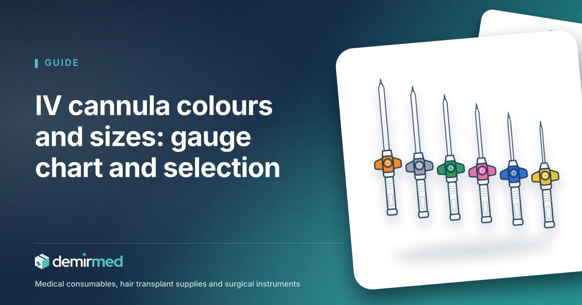
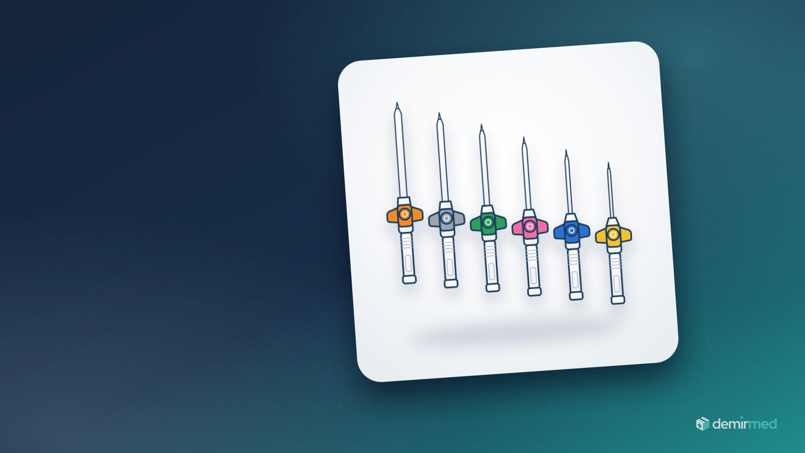
IV cannula colours are the international code (ISO 10555-5) for the gauge (G) of a peripheral IV cannula: orange 14G, grey 16G, white 17G, green 18G, pink 20G, blue 22G, yellow 24G, violet 26G. Higher numbers mean thinner cannulas and lower flow. Orange and grey suit trauma and rapid fluids, green and pink transfusion and surgery, pink and blue routine infusion, yellow and violet children and small veins.
Intraket, branül, angiocath: what is an IV cannula and what are its parts?
A peripheral venous catheter (peripheral IV cannula) is a short, flexible, single-use plastic cannula placed in a superficial vein of the arm or the back of the hand. The metal needle inside it (the stylet) guides entry into the vein; once the catheter has been advanced, the needle is withdrawn and only the flexible catheter stays in the vein. This design is called an over-the-needle catheter. The cannula provides reliable venous access for fluids, IV drugs, blood products and contrast media.
One device, many names
In general terms a cannula is any thin tube inserted into the body; in the context of venous access it means the IV cannula. Several names are used for the same device in practice, and Turkish search terms in particular mix them freely:
- Intraket: The name most often heard on Turkish wards and in emergency departments; it derives from a trade name that was once widespread.
- Branül (branula): Common in hospital jargon and product lists; it is also brand-derived.
- Anjiokat (angiocath): Used especially in theatres and emergency settings, again a term that grew out of a trade name.
- IV cannula, venous access cannula, "IV needle": Generic names. "IV needle" is widely used, but the part that stays in the vein is a plastic catheter, not a needle.
- Peripheral venous catheter (PVC, PIVC): The technical name used in guidelines and standards.
In short, "what is an intraket", "what is a branül" and "what is an angiocath" have the same answer: they all describe the peripheral IV cannula. Product pages in our IV Cannulas & Vascular Access category use IV cannula as the technical name, with branula in brackets.
Parts of an IV cannula
| Part | Function | What to check when choosing |
|---|---|---|
| Needle (stylet) | Passes through skin and vein wall and guides the catheter; the tip has a bevelled cut | Never reinserted into the catheter once withdrawn |
| Catheter | The flexible tube that stays in the vein; made of fluoropolymers such as FEP or PTFE, or of polyurethane | Polyurethane softens at body temperature; most models carry radiopaque stripes visible on X-ray |
| Flashback chamber | Clear chamber in which blood appears when the needle enters the vein | Some models also show flashback along the catheter |
| Wings | Make the cannula easier to hold and to secure to the skin | Winged and wingless models exist |
| Injection port | Valved port on top; allows bolus drugs without interrupting the infusion | Ported and non-ported models exist; keep the port cap closed |
| Luer-lock hub and cap | Threaded connection for the infusion set, extension line or syringe | Close any unused end with a sterile cap |
A ported cannula makes frequent bolus medication on wards practical. A non-ported cannula has fewer open access points; some institutions prefer the non-ported model combined with a needle-free connector.
Safety IV cannulas (needlestick protection)
A used stylet is one of the riskiest sharps for healthcare workers. In safety IV cannulas the needle tip is covered automatically by a clip as the needle is withdrawn (passive system), or the needle retracts into the barrel at the press of a button (active system). Some models also include a blood control valve that reduces blood spill from the hub after needle removal. European Council Directive 2010/32/EU requires, based on risk assessment, the provision of medical devices with safety-engineered protection mechanisms.
IV cannula colours and sizes: gauge, diameter and flow chart
What does gauge (G) mean?
Gauge is an old measuring system for wire and needle diameters, and it runs in reverse: the higher the number, the thinner the cannula. A 14G cannula has an outer diameter of about 2.1 mm, while a 24G cannula measures about 0.7 mm. Gauge refers to the outer diameter; the inner diameter (lumen) depends on wall thickness and differs slightly between manufacturers.
Why does a small change in diameter change flow so much? In laminar flow, the flow rate rises with the fourth power of the radius and falls as the cannula gets longer (Poiseuille's law). A slightly wider bore multiplies the flow, and at the same gauge a short cannula flows faster than a long one. This is why short, wide peripheral cannulas are chosen when fluids or blood must be given quickly.
ISO 10555-5 colour code chart
The colour code of over-the-needle peripheral catheters is defined in ISO 10555-5. The colour sits on the hub, wings or cap so the size can be recognised at a glance. The diameter, length and flow values below are typical ranges.
| Colour | Gauge | Outer diameter (approx.) | Typical length | Gravity flow (approx.) |
|---|---|---|---|---|
| Orange | 14G | 2.0–2.2 mm | 45–51 mm | 240–330 ml/min |
| Grey | 16G | 1.7–1.8 mm | 45–51 mm | 180–220 ml/min |
| White | 17G | 1.4–1.5 mm | 45 mm | 125–140 ml/min |
| Green | 18G | 1.2–1.3 mm | 30–45 mm | 90–105 ml/min |
| Pink | 20G | 1.0–1.1 mm | 25–32 mm | 55–65 ml/min |
| Blue | 22G | 0.8–0.9 mm | 25 mm | 30–36 ml/min |
| Yellow | 24G | 0.7 mm | 14–19 mm | 18–24 ml/min |
| Violet | 26G | 0.6 mm | 19 mm | 13–17 ml/min |
Flow values are approximate figures that manufacturers measure under standard test conditions, usually with water under gravity; short and long models also exist within the same gauge. Cannula length, the test set-up, the viscosity of the fluid (blood, colloids, contrast), the length of the infusion set, the height of the bag and the condition of the vein all change the real flow. Check the pack of the product you use for exact values.
Do not confuse them with syringe needle colours
The colour code of single-use hypodermic needles is defined in a different standard (ISO 6009). On syringe needles pink marks 18G, green 21G, blue 23G and orange 25G, so they do not match IV cannula colours. Keeping syringe needles and cannulas in separate compartments in the store room and on the treatment trolley prevents mix-ups.
IV cannula colours and their meaning: which size for which patient?
The colour code alone does not tell you which cannula is right. The flow required by the therapy, the type of fluid, the duration of treatment and the size of the vein are assessed together. The table below summarises common clinical uses; the clinical decision rests with the physician and the institution's protocol.
| Colour and gauge | Typical use | Note |
|---|---|---|
| Orange 14G, grey 16G | Major trauma, severe haemorrhage, rapid fluid and blood replacement in theatre | Needs a large, visible vein; insertion is more painful |
| White 17G | Intermediate option between 16G and 18G, rapid fluids | Not in every manufacturer's range |
| Green 18G | Blood transfusion, surgery, rapid infusion, contrast CT | The wide size most often chosen in adults |
| Pink 20G | General adult infusion, blood products, contrast imaging | Balance between flow and patient comfort |
| Blue 22G | Routine infusion and IV drugs; adults with small veins, elderly, oncology patients, older children | Among the most used sizes on adult wards |
| Yellow 24G | Paediatrics, neonates, elderly with fragile veins, oncology | Low flow; not suitable for rapid fluids |
| Violet 26G | Neonates, premature infants and very small veins | Lowest flow; not in every range |
Orange IV cannula (14G) and grey IV cannula (16G)
Orange and grey are the widest peripheral sizes. They are used in major trauma, massive haemorrhage, major surgery and rapid volume replacement; because they are short and wide, they deliver flows of several hundred millilitres per minute in these situations. They need a large, visible vein. They are not chosen for routine ward infusion without reason, because they irritate the vein wall more.
White IV cannula (17G)
17G sits between 16G and 18G and is not found in every manufacturer's range. It is used as an intermediate option when rapid fluids are needed but a 16G cannula cannot be placed.
Green IV cannula (18G)
The green cannula is 18G. In adults it is the wide size most often chosen for blood transfusion, pre-operative venous access, rapid infusion and contrast-enhanced computed tomography (CT). If it will be used with a power injector for contrast, check the maximum pressure and flow the cannula allows on its pack.
Pink IV cannula (20G)
The pink cannula is 20G. It is a versatile adult size for general infusion, antibiotics and other IV drugs, blood products and contrast imaging. It strikes a good balance between flow capacity and patient comfort. In our catalogue it is listed as the pink IV cannula 20G.
Blue IV cannula (22G)
The blue cannula is 22G. It is used for routine infusion and IV medication, in adults with small veins, the elderly, oncology patients and older children. Being small relative to the vein, it irritates the vein wall less and insertion is less painful. Blood can also be transfused through a 22G cannula; flow is slower, and the decision follows the institution's protocol. Our catalogue lists it as the blue IV cannula 22G.
Yellow IV cannula (24G)
The yellow cannula is 24G. It is preferred for infants and children, in neonatal units, for elderly patients with thin, fragile veins and for patients receiving chemotherapy. Its flow is low and it is not suitable for rapid fluid or blood replacement, although it may be used for paediatric transfusions according to protocol. In our catalogue it is the yellow IV cannula 24G.
Violet IV cannula (26G)
The violet (purple) cannula is 26G, the thinnest peripheral size in common use. It is used for neonates, premature infants and very small veins, and its flow is limited to roughly 13–17 ml per minute.
Colour code
ISO 10555-5 IV cannula colours
Choosing the right IV cannula: gauge, vein and site by situation
The core rule: the smallest gauge that meets the therapy
The common recommendation of the guidelines is to choose the smallest gauge that accommodates the prescribed therapy. A cannula that is too wide for the vein obstructs blood flow around it, irritates the inner vein wall and raises the risk of phlebitis and infiltration. On the other hand, a cannula that is too thin for a patient who needs rapid fluids or a transfusion prevents the target flow and leads to a second insertion.
Vein assessment
- Clarify the therapy: What will be given (crystalloid, blood, contrast, irritant drug), at what rate and for how many days?
- Assess the vein: Look for a soft, straight vein that refills when pressed and does not lie over a joint; skip hardened, tortuous, bruised or recently punctured areas.
- Check the cannula-to-vein fit: The cannula should not fill the vein completely; blood must be able to flow around it. Centres that use ultrasound aim for the catheter to occupy less than half of the vein diameter (a catheter-to-vein ratio of about 45% or less).
- Add patient factors: Age, dehydration, obesity, anticoagulant use, history of chemotherapy and previous difficult venous access.
- Choose gauge and length: From the range in the table, pick the smallest size that does not strain the vein.
Quick selection by situation
| Clinical situation | Commonly chosen size |
|---|---|
| Major trauma, massive haemorrhage, rapid volume | 14G–16G (two separate lines if needed) |
| Adult blood transfusion | 18G–20G; 22G if protocol allows |
| Surgery and day surgery | 18G–20G |
| Contrast CT, power injection | 18G–20G (within the manufacturer's pressure limit) |
| Routine infusion, antibiotics, IV drugs | 20G–22G |
| Fluid and drug line in hair transplant and outpatient procedures | 20G–22G |
| Elderly, thin and fragile veins, oncology | 22G–24G |
| Children | 22G–24G |
| Neonates and premature infants | 24G–26G |
General principles for site selection
- In adults, the upper extremity (forearm and back of the hand) is preferred; the lower extremity is used only when unavoidable.
- Sites over joints (antecubital fossa, wrist) let the cannula move with flexion; the forearm is better for longer infusions.
- According to institutional protocol, avoid the arm on the side of an arteriovenous fistula, lymphoedema or axillary lymph node dissection, infected, burned or damaged skin, and sites distal to a recent puncture in the same vein.
- In infants, veins of the hand, foot and, in neonates, the scalp can be used; the choice is made by a team experienced in paediatrics.
- For difficult venous access, ultrasound guidance or a more experienced clinician is preferred over repeated blind attempts.
Ported, non-ported and safety models
On wards where bolus drugs are given frequently, a ported model eases the workflow; a safety model reduces the risk of needlestick injury from the stylet.
Securement, infusion set connection, care and disposal
Securement and dressing
Good securement stops the cannula moving inside the vein, prevents dislodgement and reduces mechanical phlebitis. The insertion site is covered with a sterile transparent semipermeable dressing or a sterile gauze dressing. Transparent dressings are widely preferred for peripheral cannulas because the site can be inspected without removing the dressing; if the patient is sweating or the site is bleeding or oozing, gauze is used until this resolves. Wings and the extension line can be secured with additional tape, but tape must not cover the insertion site or block the view. Write the insertion date and the gauge on the dressing, and replace the dressing when it becomes damp, loose or soiled.
Infusion set connection and drip-rate calculation
After the cannula has been placed, a primed infusion set is screwed onto the Luer-lock hub; check that the connection is tight and that no air remains in the line. The IV infusion set in our catalogue consists of a clear drip chamber, a roller clamp and a Luer connector, and is sold in boxes of 50.
The drip rate is calculated from the set's drop factor. Gravity infusion sets made to ISO 8536-4 deliver 20 drops ≈ 1 ml; sets labelled 15 or 10 drops/ml are also on the market, and micro-drip (paediatric) sets deliver 60 drops ≈ 1 ml. The drop factor of your set is printed on its pack, and the calculation follows it:
- Formula: drops/min = volume (ml) × drop factor ÷ time (min)
- Example: 1000 ml over 8 hours with a 20 drops/ml set: 1000 × 20 ÷ 480 ≈ 42 drops/min
Blood products are not given through a standard infusion set but through a transfusion set with a filter. Infusion solutions such as isotonic saline come in single-dose bottles or bags; use, dose and rate are the physician's decision. For cannula and infusion set options, see the Vascular Access & Infusion category.
Flushing
Cannula patency is maintained by flushing with 0.9% sodium chloride before and after medication and regularly in cannulas used intermittently. Volume and frequency follow the institution's protocol; many protocols specify a 5–10 ml flush from a single-dose ampoule or a prefilled flush syringe. Because small syringes generate higher pressure for the same force, a 10 cc syringe is usually used for flushing. If you feel resistance, do not force it; treat the cannula as occluded and assess it. Large-volume infusion bottles are not used as a flush source for more than one patient. Perform hand hygiene before every contact with the cannula, port or connector (the WHO five moments) and scrub the access point with an antiseptic.
Daily checks and signs of phlebitis
Inspect and palpate the insertion site at the intervals set by the institution's protocol, before every drug administration and whenever the patient reports pain; intensive care and paediatric patients are checked more often.
- Phlebitis: Pain or tenderness, redness, warmth, swelling, a palpable cord along the vein and, at a late stage, purulent discharge. Many institutions use scales such as the VIP (Visual Infusion Phlebitis) score.
- Infiltration and extravasation: Swelling, pallor, coolness or tightness at the site, slowing flow and leakage. Extravasation of irritant or tissue-damaging drugs requires urgent assessment.
- Infection: Discharge, spreading redness, unexplained fever.
If any of these signs is present, remove the cannula, place a new sterile cannula at another site if still needed, and document the event.
How often should an IV cannula be replaced?
The CDC guideline for the prevention of intravascular catheter-related infections (2011) states that in adults there is no need to replace peripheral catheters more frequently than every 72–96 hours to reduce the risk of infection and phlebitis; in children it recommends replacement only when clinically indicated. The INS standards of practice also base removal on clinical indication rather than a fixed schedule: signs of complications, occlusion, dislodgement or completion of therapy. Follow whichever approach your institution's protocol has adopted. According to the CDC, cannulas placed in emergencies where asepsis could not be ensured are replaced as soon as possible, within 48 hours. A cannula that is no longer needed is removed without delay.
Removal and disposal
- After removing the cannula, press on the site with a sterile gauze swab until bleeding stops, then cover the point with a small sterile plaster such as our round injection plasters, or with a padded sterile dressing (adhesive wound dressing 10x10) if it oozes. Check that the catheter tip is intact.
- Discard the stylet at the point of use, without recapping, straight into a puncture-resistant sharps container. A container should be within arm's reach wherever venous access is performed.
- Fill the container up to its fill line, or to about three quarters if there is none; do not press the waste down. The full container and blood-contaminated sets, swabs and gloves are segregated and handed over under the institution's medical waste procedure and the Turkish Regulation on the Control of Medical Waste.

Selection
The smallest gauge that meets the therapy
Common mistakes with IV cannulas
- A gauge too large for the vein: Choosing a big cannula "just in case" increases phlebitis, infiltration and pain. If rapid flow is not needed, a smaller size is enough.
- A gauge too small for the need: Placing a 24G in an adult who is due for a transfusion or rapid fluids prevents the target flow and leads to a second insertion.
- Poor securement: A cannula that moves irritates the vein wall, dislodges and raises infection risk. Taping that covers the insertion site and blocks inspection is also a mistake.
- Repeated attempts with the same cannula: After a failed attempt the same cannula is not reused; open a new sterile cannula for each attempt. Reinserting a withdrawn needle into the catheter can shear the catheter and let a fragment enter the circulation. Most protocols recommend handing over to a more experienced clinician or to ultrasound guidance after two unsuccessful attempts.
- Recapping the needle: Putting the cap back on a used needle is a well-known cause of needlestick injuries. Discard the stylet straight into the sharps container; never leave it on a tray, the bed or in a pocket.
- Damaged or expired packs: A cannula in a torn, wet or unsealed pack, or one past its expiry date, is not considered sterile and is not used.
- Confusing colours with syringe needles: The needle colour code (ISO 6009) is different; on a syringe needle pink is 18G.
- Idle cannulas: A cannula left in place for days without being used is an unnecessary infection risk.
- Missing documentation: Not recording insertion date, gauge, site and number of attempts makes replacement tracking difficult.
IV cannula stocking and purchasing checklist for clinics
How many of each gauge should you keep?
The right split depends on your patient profile and past consumption. The distribution below is only an illustrative starting plan and should be updated with the real consumption of the first few months.
| Unit type | 18G green | 20G pink | 22G blue | 24G yellow |
|---|---|---|---|---|
| Adult outpatient clinic, day procedures | 5–10% | 30–40% | 45–55% | 5–10% |
| Emergency department | 20–30% | 35–45% | 25–30% | 5–10% |
| Operating theatre | 30–40% | 35–45% | 15–25% | 0–5% |
| Paediatrics | — | 5–10% | 30–40% | 50–60% |
Emergency departments and theatres also keep a small number of 14G and 16G cannulas, and units caring for neonates keep 26G.
Checklist
- Gauge range: At least 20G, 22G and 24G; 18G and wider sizes for emergency and theatre; 26G for neonates.
- Design: Ported or non-ported, winged, safety model; a single design per ward lowers the training load.
- Pack unit: Our cannulas come individually sterile-packed, 100 per box; the infusion set 50 per box.
- Sterility: Check each individual pack for the sterile symbol and sterilisation method, the single-use symbol (a crossed-out 2) and an intact seal.
- Expiry date and lot: Check on delivery and rotate stock first-expiry, first-out (FEFO). Shelf life varies by manufacturer.
- Storage: In the original box; cool, dry, out of direct sunlight, on shelves off the floor; protected from crushing and moisture.
- Reorder point: Set from lead time and monthly consumption; details in our clinic stock management guide.
- Monitoring: For patient monitoring in rooms where transfusion or sedation is performed, see our pulse oximetry guide.
- CE, ÜTS and training: Check the CE mark on the packaging and the product's ÜTS registration; plan staff training when switching to a new model, especially a safety cannula.
Venous access supplies list
A tray prepared for inserting a peripheral line usually contains:
- A sterile IV cannula of the right gauge
- Examination gloves
- Tourniquet
- The skin antiseptic specified in the institution's protocol (for example alcoholic chlorhexidine)
- Sterile gauze swabs; non-sterile gauze swabs 10x10 for skin and surface cleansing
- Sterile transparent dressing and securement tape
- A flush syringe prepared with normal saline
- If needed, a primed infusion set and infusion solution
- A sharps container

Safe disposal
Stylet straight into the sharps container
Sourcing IV cannulas and venous access supplies from Demir Med
The peripheral IV cannulas in our catalogue are listed in three sizes in the IV Cannulas & Vascular Access category:
- Blue IV cannula 22G: the mid-size for routine infusion and IV medication.
- Pink IV cannula 20G: higher flow, blood products and venous access before contrast imaging.
- Yellow IV cannula 24G: paediatric patients and thin veins.
All three are over-the-needle catheters with a clear flashback chamber and a Luer-lock hub, sterile and single-use, individually packed and sold 100 per box. The injection port and wing configuration is specified at quotation and confirmed in writing in the quotation. If you need other sizes found on the market, such as 14G, 16G, 17G, 18G or 26G, or a safety model, you can state this in your quote request.
For infusion, the IV infusion set (50 per box) is listed in the same Vascular Access & Infusion category; isotonic saline and other infusion solutions are not part of our catalogue. For aftercare, the adhesive wound dressing 10x10 (50 per box) and round injection plasters (100 per box), for safe disposal the sharps container in 1.5, 3 and 5 L sizes, and for personal protection non-sterile examination gloves in sizes S, M and L can all be added to the same quote request.
The process works like this: add products to your list with "Add to quote" and send a quote request; our sales team replies with a written quotation within 1 business day. In-stock orders confirmed before noon are delivered the same day within İstanbul and within 1–3 business days across Türkiye; the lot number and expiry date are stated on the delivery note. For bulk purchase planning see our hospital consumables purchasing guide, for sterility and packaging checks our sterile medical supplies guide, and for questions reach us through the contact page.
Sources and standards
- ISO 10555-1 and ISO 10555-5: Sterile, single-use intravascular catheters; general requirements and over-needle peripheral catheters (including the colour code)
- ISO 6009: Colour coding of single-use hypodermic needles
- ISO 80369-7: Small-bore (Luer) connectors for intravascular applications
- ISO 8536-4: Single-use, gravity-feed infusion sets
- ISO 15223-1: Symbols used on medical device labels
- CDC, Guidelines for the Prevention of Intravascular Catheter-Related Infections (2011)
- Infusion Nurses Society (INS), Infusion Therapy Standards of Practice
- WHO, Guidelines on Hand Hygiene in Health Care (2009)
- European Council Directive 2010/32/EU on the prevention of sharp injuries in the hospital and healthcare sector
- Turkish Regulation on the Control of Medical Waste (Tıbbi Atıkların Kontrolü Yönetmeliği)